
Most people who have encountered GHK-Cu know it as a skincare ingredient, the copper peptide credited with firming aging skin and accelerating wound repair. That reputation is well-earned and reasonably well-supported. But a quieter, more consequential scientific story has been developing in parallel, one that asks whether this same tripeptide might protect and even help rebuild neural tissue. The contrast is striking: decades of cosmetic research on one side, a still-early body of preclinical neuroprotection data on the other.
One detail from the basic science tends to stop researchers mid-sentence. Plasma concentrations of GHK sit around 200 ng/mL in a healthy 25-year-old and fall to roughly 80 ng/mL by age 60. That age-related decline tracks closely with reduced tissue repair capacity and rising systemic inflammation. Whether it also tracks with neural vulnerability is the question driving this emerging research frontier.
This article works through the mechanistic case for GHK-Cu neuroprotective effects research, the animal model evidence, and the gene expression data that makes this area genuinely interesting. It also holds the line on what that evidence cannot yet support: human applicability remains unproven, and the gap between preclinical promise and clinical reality is large.
What Is GHK-Cu and Why Does It Matter for the Nervous System
The story begins in 1973, when biochemist Loren Pickart isolated a small peptide fragment from human albumin that appeared to stimulate liver tissue regeneration in vitro. That fragment, glycyl-histidyl-lysine, became the basis for what is now known as GHK-Cu once its copper-binding properties were characterized. The initial discovery, published in Nature New Biology, was primarily a tissue regeneration finding, not a neural one, but it established the foundational biology that researchers would spend the next five decades unpacking.
The copper coordination chemistry is where GHK-Cu distinguishes itself from free copper ions, which are cytotoxic at equivalent concentrations. GHK binds copper(II) in a square-planar geometry, with coordination sites provided by the alpha-amino group of glycine, the imidazole nitrogen of histidine, and two deprotonated amide nitrogens from the peptide backbone. This precise geometric arrangement allows GHK-Cu to donate copper to cuproenzymes and trigger specific downstream signaling cascades without the oxidative toxicity associated with unbound Cu(II).
The age-related plasma decline adds another dimension. At age 20-25, circulating GHK measures approximately 200 ng/mL; by age 60, that figure drops to around 80 ng/mL, a reduction of roughly 60 percent. These numbers highlight why researchers began looking beyond skin biology. If GHK participates in tissue repair and gene regulation across multiple organ systems, its declining presence with age becomes a plausible contributor to neural vulnerability, not just dermal thinning.
The pivot toward neural applications followed logically from the recognition that GHK-Cu modulates overlapping pathways in wound healing and gene expression that are also relevant to nervous system maintenance. Shared mechanisms in extracellular matrix remodeling, anti-inflammatory signaling, and antioxidant enzyme regulation provided the rationale, even before neural-specific data existed.
How GHK-Cu Works at the Molecular Level
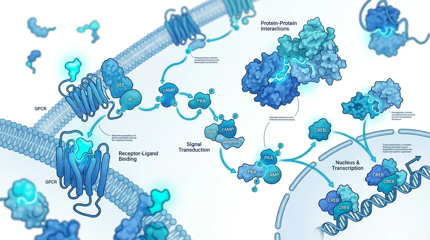
The copper binding affinity of GHK is exceptional by any standard. With a log K value of approximately 16.4, it far exceeds the binding strength of many endogenous copper-chelating agents. This tight coordination is not merely a chemical curiosity; it determines the peptide's ability to ferry copper to cuproenzymes like superoxide dismutase and ceruloplasmin, enabling specific enzymatic cascades rather than indiscriminate metal delivery.
At the gene expression level, GHK-Cu has demonstrated broad and somewhat surprising reach. In fibroblast culture studies, it upregulates genes associated with wound healing and tissue repair, including those encoding collagen I, collagen III, and decorin, while suppressing fibronectin production. Simultaneously, it downregulates pro-inflammatory cytokine pathways including TNF-alpha and IL-6 signaling. The upregulated gene categories include extracellular matrix components, growth factor mediators, and antioxidant enzymes, while the downregulated categories center on inflammatory cytokines and tissue-degrading matrix metalloproteinases. The breakdown illustrates a pattern of coordinated tissue homeostasis rather than simple stimulation of any single pathway.
The antioxidant mechanism deserves particular attention because it is frequently mischaracterized. GHK-Cu does not function as a direct free-radical scavenger. Instead, it upregulates superoxide dismutase and catalase activity, meaning the protection it offers comes through amplifying the body's own antioxidant enzyme machinery. This distinction matters for understanding both its safety profile and its likely mode of action in neural tissue, where oxidative stress plays a prominent role in injury and neurodegeneration.
The role of copper itself is confirmed by comparative studies. GHK alone, without the metal, retains partial biological activity including effects on fibroblast migration and TGF-beta modulation. The copper-bound form is consistently more potent across assays, confirming that copper coordination is central to the peptide's bioactivity, though not entirely responsible for it.
NGF and BDNF Upregulation: The Neural Mechanism in GHK-Cu Neuroprotective Effects Research
Nerve growth factor and brain-derived neurotrophic factor are the two most studied members of the neurotrophin family. NGF supports the survival and maintenance of sympathetic and sensory neurons, promotes axonal growth, and plays a key role in the peripheral nervous system's response to injury. BDNF is broadly active in the central nervous system, where it regulates synaptic plasticity, neuronal survival, and the maintenance of myelin-producing cells. Both are considered critical to functional recovery after neural injury.
The proposed mechanism by which GHK-Cu influences these pathways operates through its broader gene modulation activity. Preclinical evidence suggests that GHK-Cu upregulates NGF expression in relevant cell types and activates BDNF-associated signaling pathways, likely through the same copper-dependent transcriptional mechanisms that govern its effects in skin and connective tissue. This is not a separate neural mechanism; it appears to be an extension of the same gene regulatory activity observed across other tissue systems.
The Connectivity Map bioinformatic data from the Broad Institute adds a provocative layer to this picture. Analysis of GHK's effects on human gene expression profiles showed that it could reverse gene expression signatures characteristic of several cancer types in silico, including neuroblastoma. Gene expression reversal in this context means that GHK modulated the transcriptional activity of neuroblastoma cells in a direction opposite to their disease-associated signature, a finding that researchers interpret as evidence of pathway-level neural modulation. It is mechanistically intriguing. It is also entirely preclinical and computational.
These pathway interactions are well-characterized in cell culture models and bioinformatic analyses. They have not been confirmed in human neural tissue. That distinction is not a technicality; it is the central limitation of GHK-Cu neuroprotective effects research at this stage.
Animal Model Evidence: Spinal Cord and Nerve Crush Studies
The most direct preclinical evidence for GHK-Cu's neural effects comes from animal models of spinal cord injury and peripheral nerve crush injury. In these models, GHK-Cu administration has been associated with stimulation of axonal regrowth and promotion of myelin formation, the two structural requirements for functional neural recovery after injury.
Across injury model types, the evidence is not uniformly strong. Axonal regrowth data from nerve crush models represents the most consistently reported finding. Myelin formation outcomes have been reported but with more variable results. Functional recovery metrics, the measures that would matter most clinically, are the least well-documented in the available literature. The breakdown across these outcome categories is important for calibrating expectations accurately, as the structural findings are more robust than the functional ones. These numbers and distinctions highlight where the evidence genuinely supports mechanistic interest and where it does not yet support clinical inference.
An important contextual parallel comes from GHK-Cu's anti-fibrotic effects across other tissue systems. The same peptide that stimulates collagen production in atrophic skin also normalizes excessive collagen deposition in fibrotic liver and lung tissue. This homeostatic regulatory pattern, where GHK-Cu appears to restore appropriate tissue architecture rather than simply amplifying production, is conceptually consistent with a neural repair role. Anti-fibrotic signaling is relevant to spinal cord injury recovery, where glial scarring represents a major barrier to axonal regeneration.
Animal studies are animal studies. The translational gap between rodent nerve crush models and human spinal cord injury is substantial, involving differences in scale, anatomy, immune response, and clinical complexity. The animal data provides mechanistic plausibility and justifies further investigation. It does not constitute evidence of efficacy in humans.
What the Gene Expression Data Actually Shows
The 2018 Biomolecules review by Pickart and Margolina catalogued GHK-Cu's effects across 32 independent biological pathways. These encompass wound repair, anti-inflammation, antioxidant defense, anti-pain nociception, anti-anxiety behavior, DNA repair activation, and rhythmic gene expression. The neural and neuroprotective subset of these pathways includes anti-anxiety signaling, nociception modulation, and the neurotrophin upregulation discussed above. The distribution across pathway categories reflects how broadly this tripeptide engages biological regulation. The breakdown illustrates why GHK-Cu is considered one of the most pleiotropic tripeptides yet characterized, and also why it is genuinely difficult to evaluate comprehensively in any single clinical context.
The Broad Institute Connectivity Map analysis deserves honest framing. Gene expression reversal, as used in this context, refers to a computational finding: GHK's transcriptional signature, when mapped against disease-associated gene expression profiles, moves in an opposing direction. For neuroblastoma, this suggests that GHK modulates gene networks relevant to neural cell biology in a potentially protective direction. It is a hypothesis-generating finding, not a demonstration of therapeutic effect. Bioinformatic associations have not predicted clinical outcomes reliably across drug development history, and there is no reason to treat this data differently.
What the gene expression data does provide is a mechanistic framework for understanding why GHK-Cu might matter in neural tissue. Pathway-level data from in vitro and computational models is the appropriate foundation for designing the next generation of experiments, not the evidence base for clinical conclusions.
How Does This Compare to GHK-Cu's Wound Healing Evidence
The wound healing and skin biology evidence for GHK-Cu occupies a meaningfully different evidence tier. A 2001 double-blind, placebo-controlled randomized trial published in the Archives of Dermatology demonstrated that topical GHK-Cu significantly improved skin laxity, density, and thickness after 12 weeks compared to placebo. That is human-level controlled evidence. No equivalent trial exists for neural applications.
Neural regeneration research is operating roughly one to two evidence tiers below the skin biology findings. The underlying mechanisms overlap substantially, including collagen and extracellular matrix remodeling, anti-inflammatory signaling, and antioxidant enzyme upregulation. That mechanistic overlap is the reason neural effects are plausible. It is not evidence that they occur at clinically meaningful magnitudes in humans. Readers familiar with GHK-Cu from a wound healing or skincare context should treat the neuroprotection data as early-stage science built on shared biological logic, not as an extension of established human evidence.
The Honest State of Human Applicability
Current evidence supports the conclusion that GHK-Cu modulates gene pathways relevant to neural biology, produces neuroprotective effects in animal injury models, and upregulates neurotrophic factors associated with neural repair. Current evidence does not support the conclusion that these effects translate to meaningful neuroprotection or nerve regeneration in living humans.
Bioavailability presents a specific challenge for neural applications that exceeds the already-significant barriers for topical skin delivery. GHK-Cu has a molecular weight of approximately 403.9 Da as the copper complex and is notably hydrophilic, with a low logP value that limits passive membrane permeation. Reaching neural tissue systemically requires crossing additional biological barriers beyond the skin, and the peptide's hydrophilicity becomes an even greater obstacle in that context. Injectable routes capable of achieving systemic concentrations fall into drug territory under regulatory frameworks in the US and EU, requiring clinical trial evidence for any indication that does not currently exist.
The biohacking market's enthusiasm for injectable GHK-Cu outpaces the science by a considerable margin. Rigorous human trials for neuroprotective applications would require defined patient populations, validated neural outcome measures, pharmacokinetic data confirming target tissue exposure, and dose-response characterization. None of this infrastructure currently exists for GHK-Cu in neural indications.
Where Is This Research Heading
The most credible near-term research directions involve three areas. First, formulation improvements targeting neural delivery, including nanoparticle carriers and modified peptide analogs with better CNS penetration profiles. Second, mechanistic studies focused specifically on NGF and BDNF pathway interactions in neural cell lines and organoid models, which would strengthen or challenge the current pathway-level hypotheses. Third, neurodegenerative disease model studies in animals, particularly models of Parkinson's disease and peripheral neuropathy, where the anti-inflammatory and antioxidant mechanisms have clear theoretical relevance.
GHK-Cu belongs to a broader class of copper-binding peptides being investigated for neuroprotective properties, including AHK-Cu and various synthetic copper chelate scaffolds. Comparative data consistently shows that the specific N-terminal glycine of GHK provides optimal copper binding geometry, with alanine substitution reducing affinity by approximately one order of magnitude. This structural specificity suggests that GHK-Cu's neural effects, if confirmed, will not be easily replicated by simpler copper delivery approaches.
Following primary literature through databases like PubMed rather than commercial product claims will remain the most reliable way to track genuine progress in this area. The science is moving, but it is moving carefully.
GHK-Cu neuroprotective effects research represents one of the more intellectually honest frontier topics in peptide biology precisely because the mechanistic foundations are real and the clinical evidence is absent. NGF and BDNF upregulation, animal model findings in spinal cord and nerve crush injury, and pathway-level gene modulation data all point toward genuine biological activity in neural systems. None of it translates directly to human therapeutic conclusions. The age-related plasma decline, the copper coordination chemistry, and the breadth of gene regulatory activity make this a research area worth watching seriously. It also requires exactly the patience that distinguishes careful science from premature application. Track the primary literature. The story is still being written.
Sources
- substack.com — drtraster.substack.com
- lidsen.com — lidsen.com
- healthrx.com — healthrx.com
- mdpi.com — mdpi.com
- nih.gov — pubmed.ncbi.nlm.nih.gov
- nih.gov — pmc.ncbi.nlm.nih.gov
- researchgate.net — researchgate.net
- nih.gov — pmc.ncbi.nlm.nih.gov
- worldpharmatoday.com — worldpharmatoday.com
- medsci.org — medsci.org
- youtube.com — youtube.com
- frontiersin.org — frontiersin.org
- nih.gov — pmc.ncbi.nlm.nih.gov
- nih.gov — pmc.ncbi.nlm.nih.gov
- researchgate.net — researchgate.net
- nih.gov — pmc.ncbi.nlm.nih.gov
- biorxiv.org — biorxiv.org
- rapamycin.news — rapamycin.news
- nih.gov — pmc.ncbi.nlm.nih.gov