
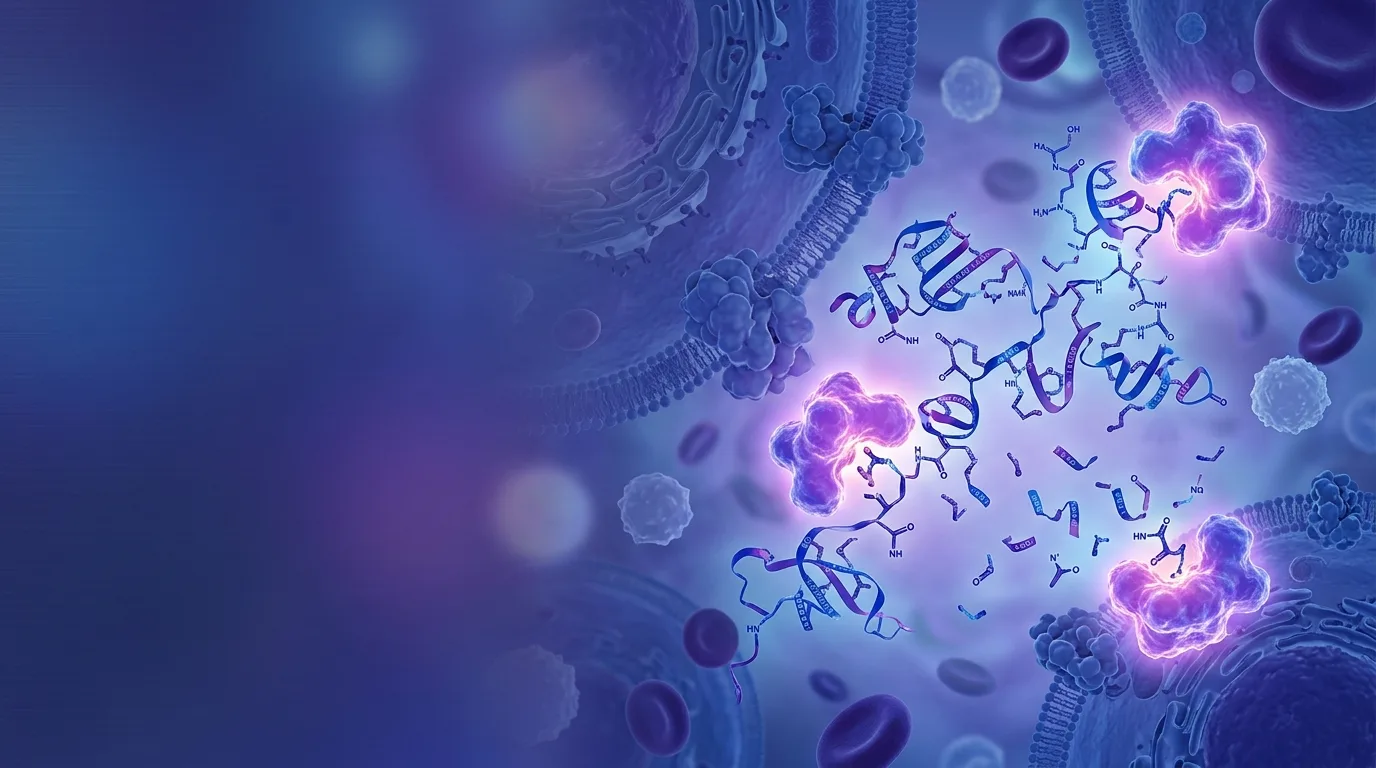
In a clinical pharmacology unit, a technician infuses a synthetic peptide into a patient's bloodstream through an IV line. The molecule has been designed, purified, and tested in animal models for months. By the time the infusion pump clicks off, roughly two minutes later, most of that peptide's biological activity is already gone. This is not a hypothetical failure. It is the ordinary fate of native glucagon-like peptide-1, or GLP-1, a hormone whose circulating half-life runs under two minutes [1]. Angiotensin II, another peptide with major roles in blood pressure regulation, fares even worse, cleared from circulation in roughly 30 seconds [2].
These numbers raise an obvious question: why do so many promising peptide-based drugs vanish almost as fast as they are administered, and what has pharmaceutical chemistry done about it? The answer sits at the intersection of biochemistry and a discipline called pharmacokinetics, the study of how drugs move through, and are eliminated from, the body. The story runs from the molecular "scissors" that dismantle peptides within moments of exposure to specific engineering fixes, D-amino acid substitution, terminal capping, and albumin binding, that have turned some peptide drugs into once-weekly injections instead of minute-by-minute infusions.
Why Therapeutic Peptides Disappear So Fast
Pharmacokinetics tracks four basic processes: absorption, distribution, metabolism, and excretion. Serum half-life, one of its central metrics, describes how long it takes for half of a drug's concentration in blood to disappear. For small-molecule drugs like aspirin, half-life is often measured in hours. For unmodified therapeutic peptides, it is frequently measured in minutes, sometimes seconds.
This is the core clinical problem. A peptide with genuine receptor-binding activity and a clean safety profile in early testing can still fail as a drug candidate if it disappears before it reaches its target tissue in sufficient concentration. Native GLP-1 clears in under two minutes. Angiotensin II clears in about 30 seconds [2]. Insulin and oxytocin, two other well-studied peptide hormones, also show short native half-lives, though their clearance depends on somewhat different physiological pathways.
Two separate clearance mechanisms drive this speed: enzymatic degradation, in which specialized proteins chemically cut the peptide apart, and renal filtration, in which the kidneys physically remove small molecules from blood [3]. Peptides are small enough to pass through the kidney's filtration membranes almost as easily as water and salt. But enzymatic degradation is often the faster and more decisive process, and it is the one this article focuses on.
Peptide Drug Metabolism: How Enzymatic Degradation Shapes Pharmacokinetics
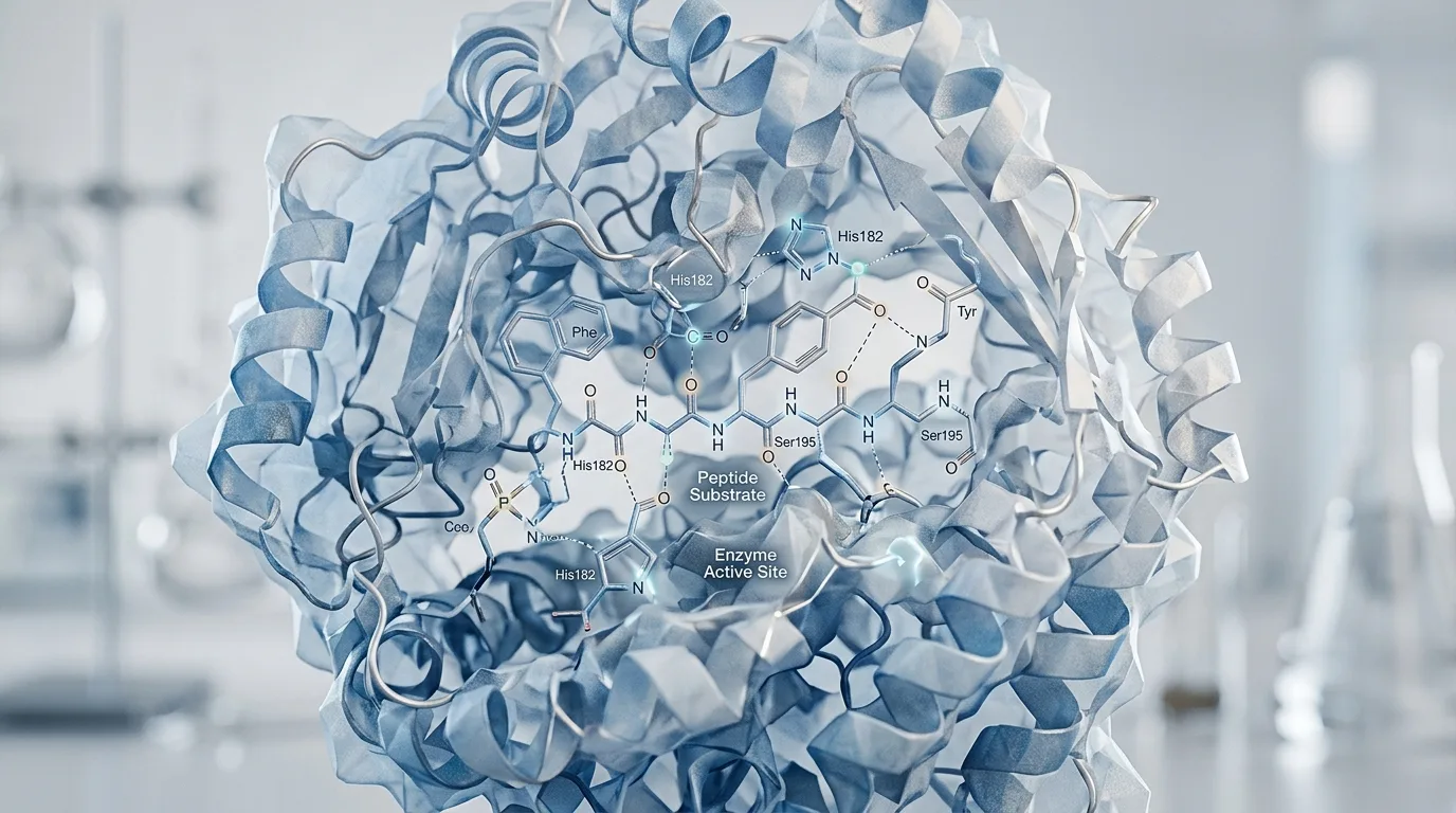
Peptidases and proteases are enzymes that target the chemical bonds holding a peptide together. Every peptide is a chain of amino acids linked by peptide bonds, and these enzymes work by cleaving those bonds [4]. The result is not simply a peptide broken into two smaller pieces sitting inertly in blood. Cleavage destroys the three-dimensional shape a peptide needs to fit into its receptor, so a single cut is often enough to eliminate biological activity entirely, even before the fragments are cleared from circulation.
The "molecular scissors" description is more than a metaphor. These enzymes have binding pockets shaped to recognize specific amino acid sequences or structural features, and they physically position the peptide bond for hydrolysis, the chemical reaction that breaks it. Different peptidases recognize different sequences, which is part of why a single peptide drug can be vulnerable to attack from multiple directions at once.
This degradation does not happen in one convenient location. Peptidases and proteases circulate in blood plasma, line the gut wall, populate liver tissue, and concentrate in the kidney, meaning a therapeutic peptide faces enzymatic threats continuously, from the moment it enters the body until it reaches, or fails to reach, its target.
Two Cutting Strategies: Exopeptidases vs Endopeptidases
Peptide-degrading enzymes fall into two broad categories based on where they cut. Exopeptidases work from the ends of the chain inward, while endopeptidases cut internally, anywhere along the sequence [5].
Picture a peptide as a strand of beads, each bead representing one amino acid. Exopeptidases behave like someone removing beads one at a time from either end of the strand. Aminopeptidases specifically target the N-terminus, the starting end of the chain, while carboxypeptidases work from the C-terminus, the opposite end [5]. Endopeptidases, by contrast, behave like scissors snapping the strand somewhere in the middle, instantly splitting one peptide into two unrelated fragments regardless of how intact the ends remain.
This distinction matters enormously for drug design, because a modification that protects one end of a peptide from aminopeptidase attack does nothing to stop an endopeptidase from cutting it in half elsewhere along the chain. Effective peptide stabilization strategies have to account for both cutting styles, not just one.
Case Study: DPP-4 and the Fast Collapse of GLP-1
Dipeptidyl peptidase-4, commonly abbreviated DPP-4, is an aminopeptidase and one of the best-documented examples of how a single enzyme can dictate a peptide drug's entire pharmacokinetic profile. DPP-4 removes exactly two amino acids from the N-terminus of GLP-1. That two-residue clip is enough to inactivate the molecule's ability to bind its receptor, and it happens fast enough that native GLP-1's half-life stays under two minutes [1].
This single enzyme-substrate relationship reshaped an entire area of pharmaceutical development. Rather than only modifying the peptide itself, researchers built a parallel drug class: DPP-4 inhibitors, commonly called gliptins, which block the enzyme rather than altering GLP-1. This is a case where the pathology of drug metabolism became the design brief for a completely separate therapeutic strategy, dipeptidyl peptidase DPP inhibitor peptide research aimed not at the hormone but at its executioner.
DPP-4's relationship with GLP-1 also illustrates something broader about how peptide drug metabolism works through enzymatic degradation and shapes overall pharmacokinetics as a field: no peptide faces a single, isolated threat. GLP-1's N-terminal vulnerability to DPP-4 is one node in a larger peptide metabolizing enzymes cascade active throughout the circulatory system, and understanding that cascade, rather than any one enzyme in isolation, is what ultimately shapes durable drug design.
Beyond One Enzyme: The Cascade of Degradation in Circulation
Few therapeutic peptides face only one enzyme. In practice, multiple peptidases act in sequence or in parallel across plasma and tissue, forming a layered degradation cascade that can dismantle a peptide through several independent routes simultaneously.
Angiotensin II offers a useful contrast to the GLP-1/DPP-4 story. Its roughly 30-second half-life [2] is not attributable to a single dominant enzyme in the way GLP-1's collapse is tied to DPP-4. Instead, angiotensin-converting enzymes and other peptidases act on it through multiple pathways nearly at once, producing an even faster clearance profile than GLP-1's despite the absence of one clean, singular mechanism.
This layered vulnerability explains a recurring frustration in peptide drug design: a modification that successfully blocks one enzyme's access point sometimes fails to meaningfully extend half-life, because a second or third peptidase simply takes over the degradation work. It is one reason combined design strategies, discussed later in this article, have become standard rather than optional. It is also worth stating plainly that the relative contribution of individual peptidases to the total clearance of many peptide drugs is not fully characterized. This remains an open question in the pharmacology literature, not a settled one.
Engineering Resistance: D-Amino Acids and Structural Substitution
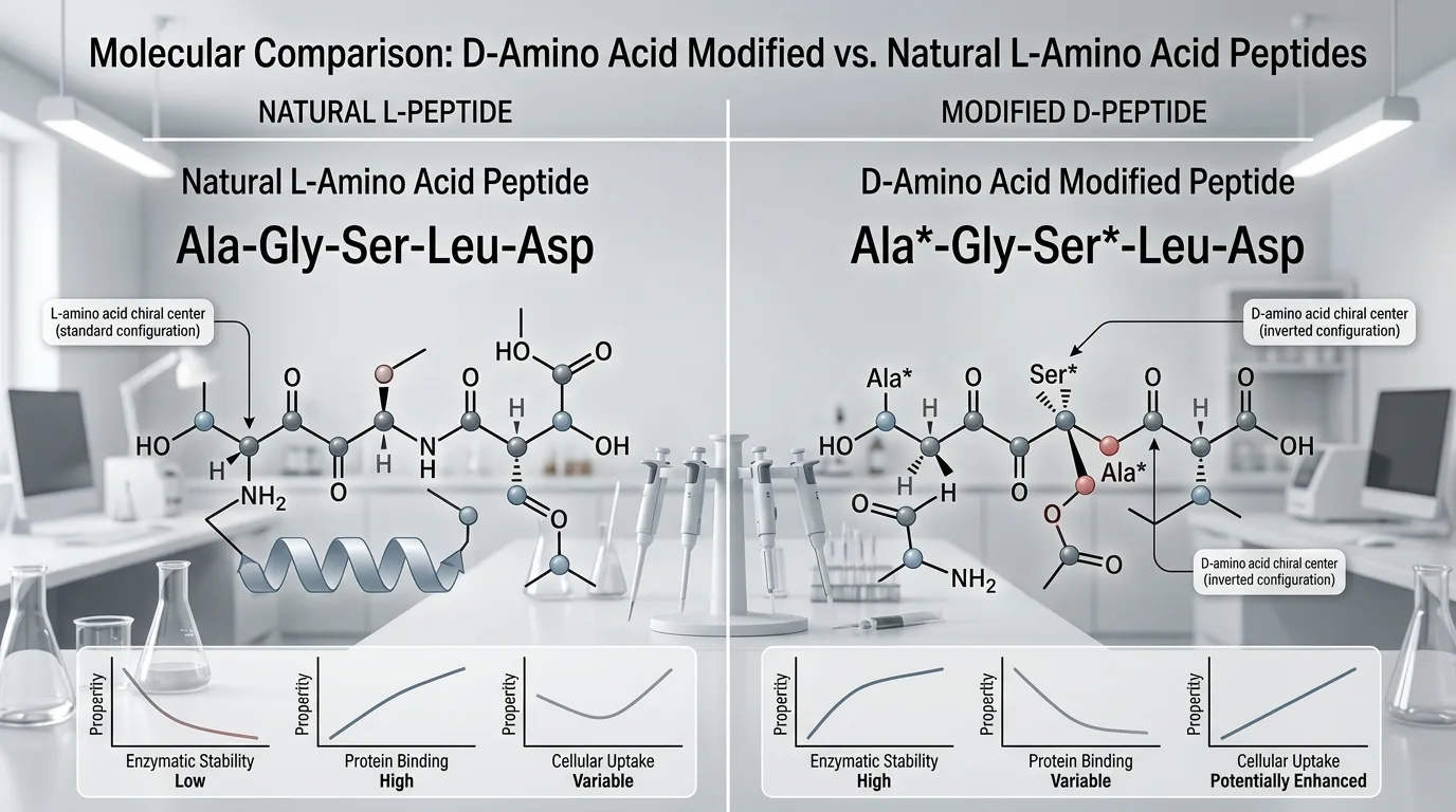
Every natural amino acid used in human proteins exists in what chemists call the L-form, one of two possible mirror-image configurations of the same molecule. The D-form is the mirror image, chemically nearly identical but spatially reversed, the way a left hand mirrors a right hand. The body's proteases evolved over millions of years to recognize and cut L-amino acid sequences almost exclusively. D-amino acids largely escape recognition [6].
This asymmetry gives peptide chemists a direct tool. Substituting specific D-amino acids at known cleavage-vulnerable positions in a peptide sequence can improve peptide metabolic stability against proteolysis, because the enzyme's binding pocket, shaped for the L-configuration, no longer fits [6]. The peptide's backbone can remain largely intact even as its individual amino acid components are swapped for their mirror-image counterparts.
The technique carries a documented caveat. D-amino acid substitution has, in some cases, been associated with potential cytotoxicity, meaning the modification is not automatically safe simply because it improves stability [7]. Each substitution has to be assessed on its own terms, peptide by peptide, position by position. This is a case-by-case engineering tool rather than a universal fix, and treating it as the latter would misrepresent what the evidence currently supports.
Capping the Ends: N-Terminal and C-Terminal Modifications
Capping refers to chemically blocking the two ends of a peptide chain so that exopeptidases have nothing to grab onto. The two most common capping methods are N-terminal acetylation, which modifies the starting end of the chain, and C-terminal amidation, which modifies the opposite end [8].
The mechanism connects directly back to the exopeptidase biology described earlier. Aminopeptidases require access to a free N-terminus to begin trimming amino acids one at a time; acetylation removes that access point. Carboxypeptidases require a free C-terminus; amidation removes that one instead [8]. In effect, capping defeats exactly the two enzyme classes that attack from the chain's outer edges.
The limitation is equally important to state. Capping does nothing to stop endopeptidases, which cut internally and never need access to either terminus in the first place. A peptide can be fully capped at both ends and still collapse within minutes if an internal cleavage site remains exposed. This is precisely why capping is rarely deployed alone in modern peptide drug design, and why it is so often paired with other stabilization strategies.
The Albumin-Binding Breakthrough and Modern Diabetes Drugs
Albumin is the most abundant protein circulating in human blood, and it has a serum half-life of its own, roughly 19 days [9]. That number alone explains why binding a short-lived peptide to albumin became one of the more consequential ideas in peptide pharmacology over the past two decades.
The protective logic works through two separate mechanisms. First, a peptide bound to albumin becomes physically shielded from the peptidases and proteases that would otherwise have direct access to its vulnerable sequences. Second, the combined peptide-albumin complex becomes far too large to pass through the kidney's filtration membranes, closing off the renal clearance route that would otherwise remove the peptide from circulation on its own [9].
The most common way to achieve this binding is through fatty-acid acylation, chemically attaching a fatty acid side chain to the peptide. That fatty acid does not bond to albumin covalently; instead, it settles into
Sources
- diabetesjournals.org -- diabetesjournals.org
- nih.gov -- pmc.ncbi.nlm.nih.gov
- nih.gov -- pmc.ncbi.nlm.nih.gov
- libretexts.org -- med.libretexts.org
- differencebetween.com -- differencebetween.com
- lifetein.com -- lifetein.com
- mdpi.com -- mdpi.com
- lifetein.com -- lifetein.com
- nih.gov -- pmc.ncbi.nlm.nih.gov