

In 2021, researchers affiliated with Lund University in Sweden began enrolling patients in a clinical trial testing exenatide, a GLP-1 receptor agonist first approved for type 2 diabetes in 2005, for signs of neuroprotection in Parkinson's disease. The trial, one of several examining the drug class outside its original metabolic context, asked a question that would have seemed strange to the endocrinologists who characterized the hormone glucagon-like peptide-1 in the gut during the 1980s. Could a molecule discovered in intestinal L-cells do something useful inside the hippocampus, a brain structure with no obvious connection to blood sugar regulation.
The Parkinson's trial built on earlier work from University College London, where neurologist Tom Foltynie and colleagues had published smaller studies on exenatide and motor function years before the Lund enrollment began. By the early 2020s, the question had spread well beyond a single lab. Pharmaceutical companies that once marketed GLP-1 drugs strictly for diabetes and, later, obesity were now funding neurology trials, a pivot with real financial stakes given that some of these companies also stand to benefit from any expanded label.
This article is not a treatment announcement. There is no approved GLP-1 receptor agonist neurodegenerative disease neuroprotection therapy, and none is imminent. What follows is an accounting of why neuroscientists, and not just endocrinologists, have started paying close attention to this drug class, grounded in receptor biology, animal models, and a long list of open questions that preclinical data alone cannot answer.
What GLP-1 Receptor Agonists Were Built to Do
Glucagon-like peptide-1 was identified in the 1980s as a hormone secreted by intestinal cells in response to food intake. Its primary job, as researchers understood it then, was straightforward: it prompts the pancreas to release insulin when blood glucose rises, while also slowing gastric emptying and reducing appetite signals in the hypothalamus.
That combination made GLP-1 an attractive target for type 2 diabetes treatment, since natural GLP-1 degrades within minutes in the bloodstream. Pharmaceutical chemists spent the 1990s and 2000s engineering longer-lasting receptor agonists, synthetic molecules that bind the same receptor but resist rapid breakdown. Exenatide, derived originally from a compound found in Gila monster saliva, became one of the first to reach the market. Later entrants, including liraglutide and semaglutide, extended the drug class into obesity treatment as well.
For most of this history, the story stayed contained within metabolism. Then researchers mapping GLP-1 receptor distribution using radiolabeling and immunohistochemistry techniques in animal tissue found something that did not fit the metabolic narrative. The receptors were not confined to the pancreas, stomach, and hypothalamus. They showed up in brain regions with no direct role in digestion or glucose handling, a finding that would eventually redirect a portion of GLP-1 research entirely.
GLP-1 Receptors in the Brain: Hippocampus and Cortex
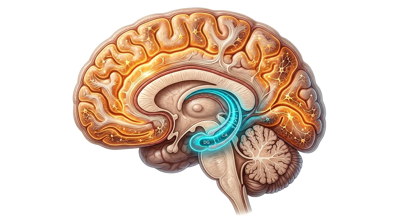
Subsequent animal studies, including work using rodent brain tissue at academic centers such as the National Institute on Aging's Intramural Research Program, identified GLP-1 receptors expressed in the hippocampus and cortex. Both regions matter enormously in neurodegenerative disease. The hippocampus is central to memory formation and is among the first structures to show measurable atrophy in Alzheimer's disease. The cortex governs executive function, planning, and much of what deteriorates in later-stage dementia.
Receptor presence alone does not prove that a drug does anything useful in that tissue. A receptor can exist in a region without that region depending on it in any meaningful way, and biological plausibility is not the same as clinical relevance. What the mapping studies did establish is that GLP-1 receptor agonists, once they cross into the central nervous system in sufficient concentration, have a physical target to act on inside memory-related circuitry.
That distinction, plausibility versus proof, runs through nearly every finding in this field. Rodent studies from multiple labs, including work associated with Christian Hölscher, a researcher who has published extensively on GLP-1 and neurodegeneration since the 2000s, described neurons in hippocampal tissue responding to GLP-1 receptor activation with changes in signaling pathways tied to cell survival. These findings reframed the drug class. GLP-1 agonists were no longer understood purely as metabolic tools; they became candidates for central nervous system study, a category shift that took roughly two decades to solidify in the literature.
The Metabolic Brain Connection: Insulin Signaling and Type 3 Diabetes
One thread connecting metabolic and neurological research involves insulin signaling inside the brain itself, separate from insulin's role in blood glucose regulation elsewhere in the body. Neurons use insulin signaling pathways for tasks related to synaptic plasticity and cell survival, and researchers studying Alzheimer's brain tissue have observed impaired insulin signaling in regions affected by the disease.
This observation led some researchers, starting roughly in the mid-2000s, to describe Alzheimer's informally as "type 3 diabetes," a term meant to capture the idea that insulin resistance might occur locally in the brain even in people without systemic diabetes. The phrase is contested. It is not a formal diagnostic category, and many researchers in the field consider it an oversimplification of a much messier picture involving multiple contributing pathologies.
Still, the hypothesis motivated a specific line of inquiry: if impaired neuronal insulin signaling contributes to Alzheimer's pathology, and GLP-1 receptor agonists influence insulin-related signaling pathways, might these drugs restore some of that signaling inside neurons. Preclinical models have shown changes consistent with this idea, but the leap from cell culture and rodent brain tissue to confirmed human mechanism has not been made. It remains a hypothesis under active investigation, not an established finding.
Neuroinflammation and Microglia: A Proposed Mechanism
A second, largely separate line of research focuses on microglia, the brain's resident immune cells. Microglia patrol brain tissue, clearing debris and responding to injury or infection. In neurodegenerative disease, however, microglia can shift into a chronic, sustained activation state, releasing inflammatory signaling molecules over months or years rather than resolving after a short defensive response.
This chronic neuroinflammation is a hypothesized contributor to disease progression in both Alzheimer's and Parkinson's models, though the exact relationship between inflammation and neuronal death remains an open question in the field. Some researchers view inflammation as a downstream consequence of protein aggregation; others view it as an active driver of further neuronal damage. The two positions are not mutually exclusive, and much of the current research tries to untangle which comes first in a given disease stage.
Preclinical studies using rodent models and cultured microglial cells have reported that GLP-1 receptor agonists appear to modulate microglial activity, shifting cells away from the sustained inflammatory state and toward a more regulated response. Cell culture work has shown reduced release of specific inflammatory signaling molecules following GLP-1 receptor activation. None of this modulation has been confirmed in living human brain tissue. Studying microglial activity directly in humans requires either postmortem tissue or advanced imaging techniques that are still being refined, which means this entire mechanism rests on animal and cell-based evidence for now.
Protein Aggregation: Amyloid-Beta, Tau, and Alpha-Synuclein
Alzheimer's disease is characterized, in its classical pathological description, by two protein abnormalities: amyloid-beta plaques that accumulate between neurons, and tau tangles that form inside neurons when a normally stabilizing protein misfolds and clumps together. Parkinson's disease involves a different protein, alpha-synuclein, which aggregates into structures called Lewy bodies inside affected neurons, particularly in the substantia nigra, a region involved in movement control.
Preclinical studies in mouse models genetically engineered to develop amyloid plaques have reported reduced plaque accumulation following GLP-1 receptor agonist exposure. Similar studies in Parkinson's-model animals, often using rodents exposed to neurotoxins that mimic dopaminergic neuron loss, have reported reduced alpha-synuclein aggregation alongside improved motor performance on standardized rodent behavioral tests.
The following summarizes reported directional findings across preclinical animal models, though it should be read as illustrative of a body of early research rather than a settled consensus: Alzheimer's mouse models have shown reduced amyloid-beta accumulation in several published studies; tau pathology reduction has been reported less consistently across models; Parkinson's rodent models have shown reduced alpha-synuclein aggregation alongside motor improvement in multiple independent studies.
These numbers highlight a pattern common in early-stage neurodegeneration research: findings are more consistent for some endpoints than others, and the underlying mechanism connecting reduced protein aggregation to improved neuronal survival is not fully understood. Researchers have proposed several explanations, including enhanced cellular clearance mechanisms and reduced oxidative damage, but no single pathway has been confirmed as the primary driver.
Oxidative Stress and Cellular Antioxidant Defenses
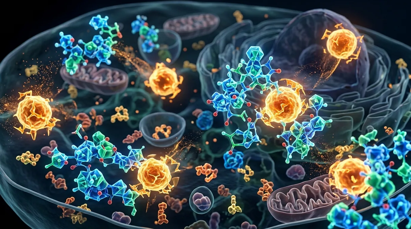
Oxidative stress describes a cellular imbalance in which reactive oxygen species, byproducts of normal metabolism that become harmful in excess, accumulate faster than a cell's antioxidant systems can neutralize them. Over time, this imbalance damages proteins, lipids, and DNA, and neurons are considered particularly vulnerable given their high metabolic demand and limited capacity for regeneration.
Oxidative damage is theorized to contribute to neuronal death across multiple neurodegenerative conditions, working alongside protein aggregation and neuroinflammation rather than as an isolated cause. Laboratory studies using cultured neuronal cells have reported that GLP-1 receptor agonists enhance the activity of specific antioxidant enzymes and reduce measurable markers of oxidative damage under experimental stress conditions.
These findings come almost entirely from in vitro cell culture experiments and rodent models exposed to induced oxidative stress. Whether the same enhancement of antioxidant defenses occurs in living human neurons, under the far more complicated conditions of an aging human brain, has not been established. The gap between a petri dish and a functioning brain is one of the largest sources of uncertainty in this entire body of research.
What Human Clinical Trials Actually Show So Far
Clinical trials evaluating GLP-1 receptor agonists for Alzheimer's and Parkinson's disease are underway at