
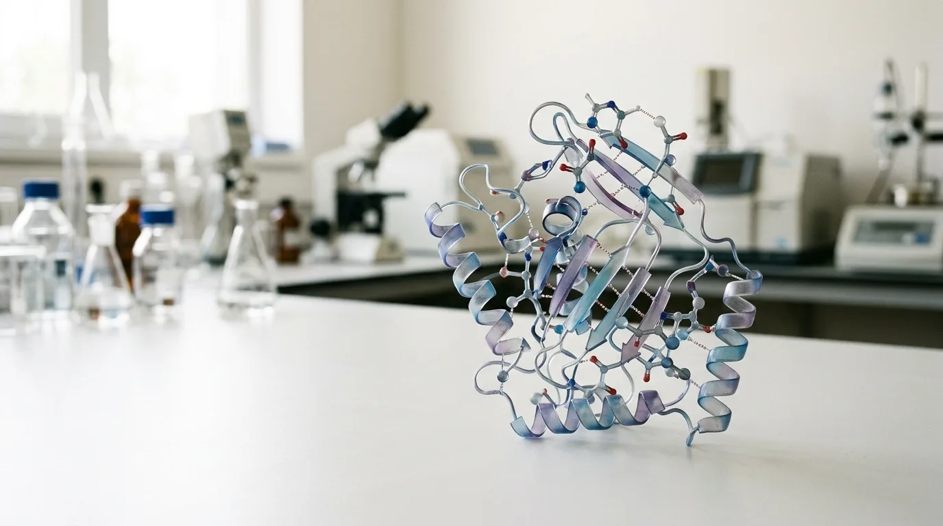
Two peptides can share an identical amino acid sequence and still behave as entirely different molecules once introduced into a biological system. One binds its target receptor with measurable precision. The other misses entirely. The difference between them is not a matter of chemistry in the conventional sense. It is geometry. This distinction sits at the heart of peptide conformational stability and three-dimensional structure, where even subtle variations in how a peptide folds determine its therapeutic efficacy.
This counterintuitive reality sits at the center of modern peptide research. A sequence of amino acids is, in the strictest sense, a set of instructions. The three-dimensional architecture that assembles from those instructions is the actual functional entity. Change the shape, even slightly, and the molecule changes its behavior. The sequence stays the same. The function does not.
BPC-157, a 15-amino-acid linear peptide that has drawn sustained attention from researchers in gastroenterology and tissue physiology, offers an early illustration of this principle. Unlike many pharmacologically studied peptides, BPC-157 carries no globally rigid folded structure. Yet it demonstrates an unusual resistance to degradation under conditions that destroy structurally more elaborate molecules. Understanding why requires a closer look at how shape emerges from sequence in the first place.
The central questions this article addresses are the following. How does three-dimensional conformation arise from a one-dimensional sequence? Why does peptide conformational stability erode under some physiological conditions and persist under others? And what tools do researchers use to predict and confirm the shapes that matter for therapeutic efficacy?
From Sequence to Shape: The Folding Problem
In 1973, Christian Anfinsen published a paper in Science that articulated a principle biochemists had been circling for years. The amino acid sequence of a protein, Anfinsen argued, encodes all the information necessary to reach the molecule's final folded conformation. No external template is required. The instructions are internal.
The thermodynamic logic follows directly. A peptide does not fold randomly and then select a shape. It samples available conformations and settles into the one with the lowest overall free energy. That state is stabilized by a network of non-covalent interactions: hydrogen bonds between backbone atoms, hydrophobic clustering of nonpolar side chains away from water, and ionic interactions between charged residues.
For short peptides, typically those fewer than 50 residues, this landscape behaves differently than it does for full proteins. The folding energy landscape is shallower. There are fewer stabilizing contacts available, which makes conformational stability both harder to achieve and more consequential for function. A large protein may tolerate the loss of a few stabilizing interactions without collapsing into an inactive form. A short peptide often cannot.
An important clarification follows from this. The folded state is rarely a single rigid structure. It is a population of interconverting conformations. The biologically relevant state is the one most heavily populated under physiological conditions, the conformation the molecule occupies most often when it encounters its target. Peptide conformational stability, in this sense, means not absolute rigidity but a meaningful bias toward the active geometry.
Alpha Helices, Beta Sheets, and Peptide Conformational Stability Three Dimensional Structure Therapeutic Efficacy
Secondary structure elements are the repeating architectural patterns that emerge from hydrogen bonding between backbone amide and carbonyl groups. They are not determined by side-chain chemistry. As Berg, Tymoczko, and Stryer established in their foundational biochemistry text, these patterns arise from the backbone geometry itself.
The alpha helix arranges residues in a right-handed spiral, with 3.6 residues per turn and a rise of 0.15 nanometers per residue. Hydrogen bonds form between the carbonyl oxygen of one residue and the amide nitrogen four residues further along the chain. The beta sheet arranges extended strands side by side, with hydrogen bonds forming between strands rather than within a single chain.
These motifs function as scaffolds. Their significance for therapeutic efficacy lies in what they accomplish spatially: they orient amino acid side chains in precise three-dimensional arrangements. Whether a peptide can physically engage a receptor binding site depends on whether those side chains arrive in the correct geometry. A helix positions its side chains along a helical path. A sheet arrays them in a planar arrangement. Neither is inherently superior; the receptor determines which is required. Peptide conformational stability in the context of three-dimensional structure directly shapes the therapeutic efficacy of these molecules.
The practical consequence appears in structure-activity relationship studies. According to a review in Trends in Pharmacological Sciences, a single amino acid substitution that disrupts a helix or sheet can abolish binding affinity entirely. The sequence changes by one unit. The shape changes at the relevant position. The functional outcome changes completely. This is not a marginal effect. It is the difference between a molecule that binds and one that does not.
Alpha helix characteristics include 3.6 residues per turn, 0.54 nm pitch, and intrachain hydrogen bonding. Beta sheets show 2 residues per repeat, 0.35 nm rise per residue, and interchain hydrogen bonding. These numbers reflect structural differences that have direct consequences for how each motif presents side chains to a receptor surface. The breakdown illustrates why even subtle changes in secondary structure content shift the functional profile of a peptide substantially.
BPC-157 and the Puzzle of Linear Stability
BPC-157 presents researchers with a structural paradox. The peptide is linear, meaning it lacks the disulfide bridges, cyclizations, or constrained geometries that typically confer stability on short sequences. By standard predictions, it should be conformationally labile and enzymatically vulnerable. Under gastric acid conditions, most linear peptides of comparable length degrade rapidly.
BPC-157 does not behave this way. Its documented stability in gastric environments has attracted repeated attention in the literature, precisely because it violates the expectation that conformational flexibility and enzymatic vulnerability travel together.
NMR studies, cited in the Journal of Physiology and Pharmacology, suggest a partial explanation. BPC-157 adopts localized turn-like conformations. These are transient but recurring structural motifs, compact arrangements of three to four residues that the peptide populates repeatedly in solution. They are not a globally folded structure. They are local geometries that emerge and dissolve on short timescales.
Whether those turn conformations constitute the peptide's functionally active state remains an open question. It is possible that the recurring turns are the structural feature responsible for the activity documented in animal models. It is equally possible that they are a structural coincidence, a property of the sequence that does not correspond causally to the observed biology. The published literature has not resolved this distinction. The question remains open, and several labs continue to study it.
Why Conformational Stability Matters: The Key Analogy
A useful analogy appears repeatedly in the peptide engineering literature. A key that deforms before reaching the lock cannot open it, regardless of whether the original cut was correct. A peptide that collapses into an inactive conformation before reaching its receptor cannot bind, regardless of whether its sequence encodes the correct pharmacophore.
Natural peptide flexibility is a double-edged property. According to the Journal of Medicinal Chemistry, flexibility allows induced-fit binding, the process by which a peptide adjusts its geometry slightly to complement the shape of a receptor binding site. This adaptive capacity is often beneficial. It extends the range of targets a peptide can engage.
The same flexibility, however, exposes the peptide backbone to protease recognition. Proteases cleave peptide bonds at specific sequence and shape signatures. A conformationally dynamic peptide presents more of those signatures more often. Conformational stability, paradoxically, can be protective not because it makes the peptide rigid but because it limits the conformations the peptide samples, reducing the fraction of time it spends in protease-accessible geometries.
Cyclization is the primary engineering strategy for addressing this problem. Constraining the peptide's backbone geometry through a covalent bond between the N-terminus and C-terminus, or between side chains, reduces conformational entropy and narrows the accessible conformational space. According to Drug Discovery Today, cyclization consistently extends half-life compared to linear counterparts of the same sequence. Across multiple reported studies, cyclized peptides show half-life extensions ranging from roughly 2-fold to more than 10-fold depending on the sequence and cyclization site. These numbers highlight why conformational engineering has become central to peptide drug design programs. The increases are not marginal; they reflect a structural change with direct pharmacokinetic consequences.
Predicting Structure Before Synthesis: Computational Tools
Before a peptide is synthesized, computational prediction tools allow researchers to generate structural hypotheses at low cost. Two dominant paradigms define the field, according to a review in Current Opinion in Structural Biology.
Ab initio methods, represented by programs such as PEP-FOLD and Rosetta, model structure from physical principles alone. They make no assumption about structural similarity to known molecules. Instead, they calculate the energy of each possible backbone configuration and identify low-energy conformations through sampling algorithms. Template-based methods, of which I-TASSER is a prominent example, anchor predictions to known homologous structures deposited in the Protein Data Bank, using sequence similarity to identify likely structural templates.
The practical workflow a researcher follows is relatively consistent across both paradigms. A sequence is entered as input. The program generates an ensemble of predicted conformations. Those conformations are ranked by calculated free energy. The lowest-energy model becomes the working structural hypothesis for subsequent experimental design.
The limitations of this workflow for short, flexible peptides are well documented. Energy landscapes for peptides fewer than 30 residues are flatter than for full proteins, meaning multiple conformations have similar predicted energies. Prediction confidence intervals are wider. The programs were developed and validated primarily on longer sequences with more stabilizing contacts, and their performance on short flexible peptides is less reliable.
Despite these limitations, computational prediction has meaningfully accelerated early-stage structure-activity relationship screening. A researcher can test dozens of virtual amino acid substitutions before committing to synthesis, identifying which changes are predicted to disrupt the active conformation and which are predicted to be tolerated. The reduction in experimental cost and time is substantial, even accounting for the irreducible need for experimental validation.
Validating the Predicted Shape in the Laboratory
Computational predictions require experimental confirmation. Three methods dominate the validation workflow, each operating at a different level of structural resolution.
Circular dichroism spectroscopy is the first-line tool. CD instruments pass polarized light through a peptide solution and measure the wavelength-dependent absorption difference between left and right circular polarization. Alpha-helical conformations produce a characteristic double-minimum spectrum at roughly 208 and 222 nanometers. Beta sheets produce a distinct single minimum near 218 nanometers. Disordered conformations produce a negative band near 200 nanometers. According to Nature Protocols, CD can quantify the approximate percentage composition of each secondary structure type, providing a rapid and relatively inexpensive first assessment of whether the predicted conformation is present in solution.
NMR spectroscopy operates at the next level of resolution. By measuring the magnetic behavior of individual atomic nuclei in the peptide, NMR resolves inter-atomic distances and backbone dihedral angles. These measurements are used to calculate a solution-state structural ensemble, a collection of atomic coordinates that collectively represent the conformational population the peptide occupies in solution. NMR captures the dynamic ensemble directly, which makes it particularly appropriate for short, flexible peptides that do not adopt a single stable conformation.
X-ray crystallography produces the highest spatial resolution available. A crystallized peptide diffracts X-rays in a pattern that, once processed, yields atomic coordinates with precision measured in fractions of a nanometer. The limitation is meaningful: crystal-packing forces, the molecular contacts imposed by the crystal lattice itself, may stabilize conformations that do not predominate in free solution. A crystal structure is not automatically equivalent to the biologically relevant solution conformation. The two methods are therefore complementary rather than redundant.
Structure-Activity Relationships and the Cost of a Single Substitution
Systematic amino acid substitution studies map which residues within a sequence are essential for maintaining the active conformation and which tolerate variation without functional consequence. This is the foundational logic of structure-activity relationship research in peptide drug design.
BPC-157 provides a concrete case. Documented studies comparing the full 15-residue sequence to truncated or substituted variants have observed sharp reductions in activity in animal models when specific residues are altered. Those reductions point to positional dependencies, specific sites within the sequence where the amino acid identity is essential for maintaining the turn conformations or other structural features associated with activity. Potency values across a substitution series, measured at each position in the sequence, show a pattern that reflects the conformational sensitivity of individual sites. The pattern is chartable: positions essential to the active conformation show steep drops in observed activity upon substitution; tolerant positions do not. The structure of that data makes the geometry-function relationship visible directly.
A critical qualification applies here. SAR findings generated in animal models do not automatically translate to equivalent conclusions in human systems. The published literature on BPC-157 and related peptides makes this distinction inconsistently, and the inconsistency is worth naming explicitly. A substitution that abolishes activity in a rodent model at a specific receptor may behave differently in a human receptor context, where binding site geometry may differ. The animal-model findings are informative; they are not determinative.
The Shape Behind the Sequence
A peptide's therapeutic relevance cannot be separated from its three-dimensional geometry. The sequence is the instruction set. The conformation is the machine that either engages its target or fails to.
Researchers studying peptides like BPC-157 have found that even structurally unassuming linear sequences can maintain functionally relevant conformations under conditions that would degrade architecturally more complex molecules. The mechanism behind that stability remains incompletely understood, which is itself a significant finding: the relationship between sequence and conformational persistence is not fully predictable from first principles in short peptides.
Computational tools, including PEP-FOLD, Rosetta, and I-TASSER, have shortened the distance between sequence design and structural hypothesis. They allow researchers to screen virtual substitutions before committing resources to synthesis. That acceleration is genuine, and it has changed the pace of early-stage peptide research. But the tools are not a substitute for experimental confirmation. CD spectroscopy, NMR, and crystallography each reveal a different layer of structural reality, and the layers do not always agree.
The field's deeper open question is the one the literature addresses least consistently. How reliably do conformational findings in animal models anticipate receptor interactions in human systems? The published record on this question is thin. It is a gap that high-resolution structural studies and more rigorous clinical translation frameworks will need to close. Until that gap narrows, the relationship between peptide conformational stability, three-dimensional structure, and therapeutic efficacy in humans remains, at its most honest level of description, a working hypothesis supported by animal evidence and awaiting harder confirmation.
Stay current with peptide structure research. Molecule Notes publishes in-depth analysis of peptide chemistry, conformational biology, and synthesis methodology. Subscribe to the newsletter to receive new articles, research summaries, and upcoming webinar announcements directly to your inbox.
Sources
- nih.gov — pmc.ncbi.nlm.nih.gov
- iptonline.com — iptonline.com
- nih.gov — pubmed.ncbi.nlm.nih.gov
- nih.gov — pmc.ncbi.nlm.nih.gov
- creative-peptides.com — creative-peptides.com
- mdpi.com — mdpi.com
- mdpi.com — mdpi.com
- acs.org — pubs.acs.org
- drugdiscoverynews.com — drugdiscoverynews.com
- nebulabio.tech — nebulabio.tech
- nih.gov — pmc.ncbi.nlm.nih.gov
- nih.gov — pmc.ncbi.nlm.nih.gov
- forbes.com — forbes.com
- cbsnews.com — cbsnews.com