
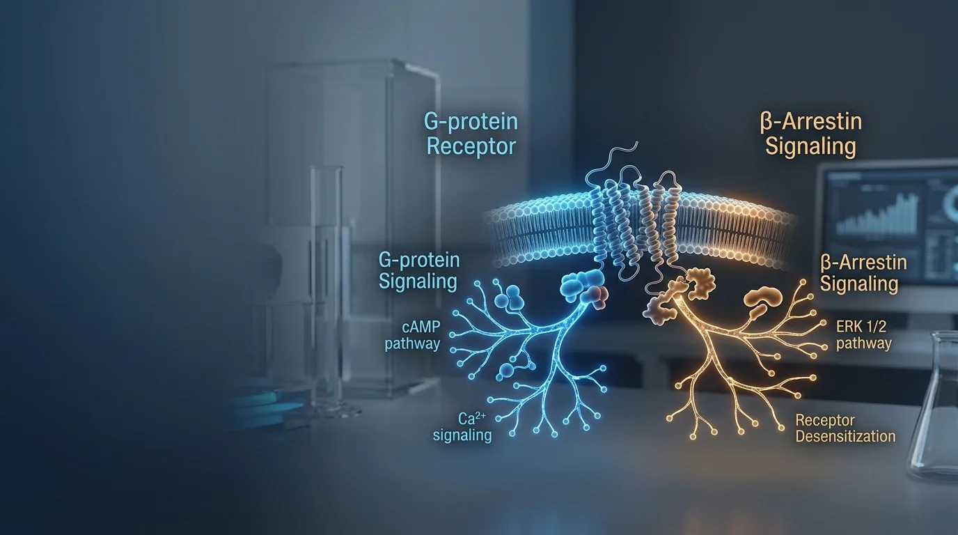
In May 2022, the FDA approved tirzepatide for type 2 diabetes, a molecule that would later become one of the best-selling obesity drugs in history under the brand name Zepbound. What drew less public attention was a parallel stream of structural biology papers, several from research groups at the University of Michigan and from Eli Lilly's own discovery teams, mapping precisely how tirzepatide grips the GLP-1 receptor compared to the body's native hormone [1]. The molecules looked similar enough on paper. Inside a cell, they behaved differently, activating different receptor pathways through peptide biased signaling that enabled selective pathway activation at the receptor level.
That difference sits at the center of a question pharmacologists have been chasing since Robert Lefkowitz's laboratory at Duke University identified a second, independent signaling role for a protein called beta-arrestin, a discovery that helped earn Lefkowitz a share of the 2012 Nobel Prize in Chemistry [2]. Two peptides can bind the same receptor, in the same pocket, and still produce different cellular outcomes downstream. Why would a small chemical substitution change which internal machinery a receptor switches on, and why would that matter for something as concrete as a side effect profile.
The answer involves a concept researchers call biased signaling, or functional selectivity, and it is reshaping how peptide drugs get designed.
What Biased Signaling Actually Means
Most peptide drugs act on a receptor family called G protein-coupled receptors, or GPCRs, proteins embedded in the cell membrane that relay signals from outside the cell to machinery inside it. A ligand is any molecule, natural or synthetic, that binds to a receptor. An agonist is a ligand that binds and activates the receptor, triggering some downstream response rather than simply occupying space.
For decades, the working assumption in pharmacology was straightforward: a receptor functioned like a single switch. Any agonist that bound and activated it would flip that switch the same way, producing a uniform signal regardless of which molecule did the binding. Potency and affinity, meaning how strongly and how tightly a ligand binds, were treated as the only variables that mattered.
Structural and cellular studies over the past two decades have undermined that assumption. A receptor is better understood as a structure capable of opening different intracellular doors, depending on which key is inserted and how that key sits in the lock. Researchers now describe this property as functional selectivity, a term that specifically distinguishes it from simple potency differences [2] [1]. Two full agonists can bind the same receptor with similar strength and still activate entirely different combinations of downstream pathways. That distinction, once treated as a laboratory curiosity, has become a design target in its own right.
G-Protein Signaling vs Beta-Arrestin: Peptide Biased Signaling and Selective Pathway Activation at the Receptor
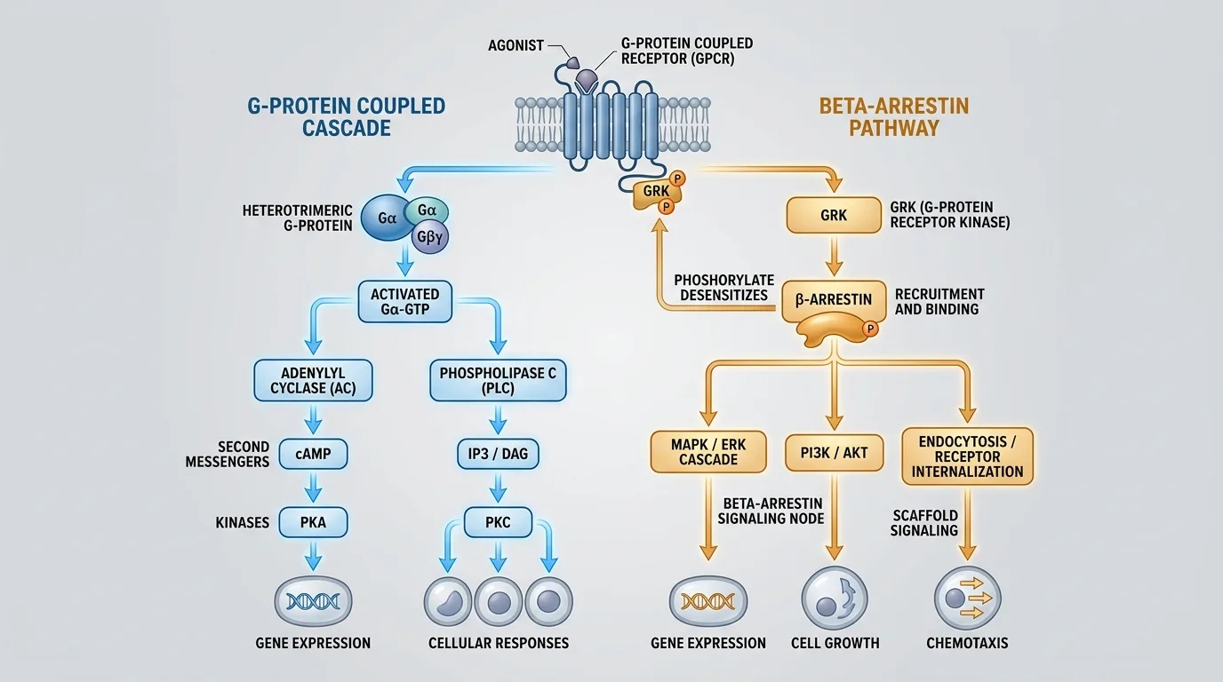
Inside the cell, an activated GPCR typically has two major routes available to it. The first is the G-protein pathway, the classical route associated with a receptor's primary, intended signal, such as the production of cyclic AMP, a small molecule that triggers cascades tied to metabolism, secretion, or muscle activity depending on the tissue.
The second route runs through beta-arrestin, a protein first identified for a much quieter job: shutting the receptor down. Beta-arrestin was originally characterized as a desensitizing agent, binding to an activated receptor, pulling it away from the cell surface through internalization, and ending the G-protein signal before it ran unchecked. Lefkowitz's Duke laboratory later found something unexpected. Beta-arrestin did not simply end signaling. It initiated its own separate signaling cascade, entirely independent of G-protein activity [2]. That discovery reframed beta-arrestin from a cleanup protein into a second messenger system in its own right.
None of this makes beta-arrestin signaling inherently harmful. Its consequences are context-dependent. In some receptor systems, beta-arrestin recruitment produces therapeutic effects; in others, it correlates with the very side effects a drug developer is trying to avoid. The pathway itself is neutral. What matters is which receptor system it operates in, and what tissue it operates on.
How a Peptide 'Chooses' a Pathway at the Molecular Level
Receptors are not rigid locks with a single correct key shape. Structural biology has shown that GPCRs are flexible proteins capable of adopting multiple three-dimensional conformations, shifting shape slightly depending on what is bound to them and how.
Peptide ligands exploit that flexibility. A specific peptide sequence can stabilize one particular conformation of the receptor over others, effectively holding the protein in a shape that favors one signaling partner. Much of the recent structural work on the GLP-1 receptor has focused on a specific region called extracellular loop 3, or ECL3, a segment that sits near the peptide-binding pocket and appears to play an outsized role in determining which intracellular partner the receptor recruits [1] [3]. Small differences in how a peptide contacts ECL3 appear to shift the receptor's internal architecture just enough to favor G-protein coupling over beta-arrestin recruitment, or the reverse.
This remains an active area of structural biology, not a settled mechanism. Researchers are still working out how generalizable ECL3-driven bias is across the broader GPCR family, versus how specific it is to the GLP-1 receptor and its close relatives. Cryo-electron microscopy studies published in recent years have offered increasingly detailed snapshots of these conformational states, but translating a static structural snapshot into a full account of cellular behavior is a harder problem than capturing the image itself [1].
GLP-1 Receptor Bias: The Central Case Study
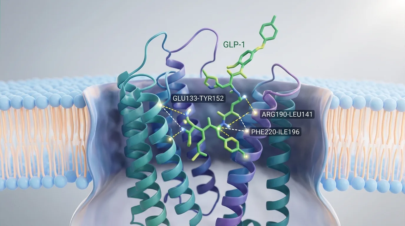
The GLP-1 receptor is one of the most closely studied GPCRs in clinical medicine, serving as the target for a class of drugs used in type 2 diabetes and, more recently, obesity. Native GLP-1, the hormone the body produces naturally, activates both the G-protein and beta-arrestin pathways at roughly comparable levels. Researchers generally treat this balanced activation profile as the baseline against which synthetic analogs are measured [1] [4]. Tirzepatide departs from that baseline. Laboratory assays characterize it as a G-protein-biased agonist at the GLP-1 receptor: a full agonist for cAMP production, the hallmark of G-protein signaling, while producing comparatively minimal beta-arrestin recruitment [1]. Semaglutide, another major GLP-1 receptor agonist, sits in a different position on that same spectrum, with a bias profile that differs from both native GLP-1 and tirzepatide, according to published receptor assays [1] [3].
Expressed roughly, published in vitro assays position native GLP-1 near a balanced midpoint between G-protein and beta-arrestin activation, semaglutide showing a partial shift toward G-protein signaling, and tirzepatide showing a more pronounced shift toward G-protein signaling with markedly reduced beta-arrestin recruitment [1] [3]. The comparison illustrates a spectrum of bias rather than a binary switch, with each molecule occupying a different point along it.
Why This Chemistry Might Explain Real-World Outcomes
The hypothesis researchers are testing runs like this: because beta-arrestin recruitment drives receptor internalization and desensitization, a G-protein-biased agonist that recruits less beta-arrestin should, in principle, produce a more sustained signal at the receptor before it gets pulled from the cell surface and shut down.
The published mechanism is hypothesized to be that tirzepatide's reduced beta-arrestin engagement allows for longer, less interrupted G-protein signaling at the GLP-1 receptor, and that this sustained signaling contributes to its observed performance in blood sugar control and weight loss in clinical trials [1] [3]. That is a hypothesis under active investigation, not a closed case. Correlation between a receptor-level bias measurement in a cell assay and a clinical outcome measured in thousands of patients is a considerable inferential leap, and researchers studying the GLP-1 receptor are careful to note that other factors, including tirzepatide's dual action at the GIP receptor, complicate any clean attribution to bias alone [1].
The pattern is not unique to GLP-1 biology. The opioid receptor system offers a second, well-documented illustration of the same design logic. Researchers have spent years trying to develop G-protein-biased opioid agonists specifically to separate pain-signaling, thought to run largely through G-protein pathways, from respiratory depression, a dangerous side effect associated with beta-arrestin recruitment at the mu-opioid receptor [2] [5]. That effort has produced mixed results in clinical testing, a caution worth keeping in mind when evaluating similarly optimistic claims made about GLP-1 receptor bias.
The Push for Biased Agonists as a Drug Design Strategy
Pharmaceutical and academic laboratories increasingly screen peptide analogs for their bias profile as a design parameter in its own right, not merely as a secondary characteristic behind potency and half-life. The appeal is straightforward on paper: amplify the therapeutic G-protein signal, minimize the beta-arrestin pathway associated with adverse effects, and end up with a molecule that does more of what a drug is supposed to do and less of what it is not.
Achieving that cleanly is difficult in practice. Most peptide analogs show partial bias rather than an absolute preference for one pathway. A molecule rarely shuts one pathway off entirely; it more commonly shifts the ratio between the two, and the degree of that shift can vary depending on which cell type, which assay system, and which readout a laboratory uses to measure it [3] [6].
There is also open skepticism within the field about how much of the observed clinical difference between drugs like tirzepatide and semaglutide can be attributed to receptor bias specifically, as opposed to differences in half-life, tissue distribution, dosing, or, in tirzepatide's case, its additional activity at the GIP receptor. Several researchers interviewed in review literature caution against treating bias as a complete explanation until more direct evidence connects receptor-level signaling data to patient-level outcomes [3] [6].
Open Questions in Biased Signaling Research
Several problems remain unresolved. Chief among them is whether bias measured in a cultured cell assay, using overexpressed receptors and engineered reporter systems, predicts how a receptor will actually behave in human tissue, where expression levels, cell type, and local signaling context differ substantially from a laboratory dish [5] [6].
A second problem is standardization. Different laboratories use different assay platforms, different reference ligands, and different mathematical models to calculate a bias factor, which makes direct comparison across published studies difficult. A molecule described as strongly biased in one lab's assay system may show a more modest bias profile when tested under another lab's conditions [3] [6].
Most of the data underlying current bias claims, for GLP-1 receptor agonists and for other GPCR systems alike, comes from in vitro cell assays and animal models. Direct mechanistic confirmation in human tissue remains limited, and researchers are candid that this gap represents one of the field's largest open questions [5] [6]. Biased signaling is an active, unsettled area of receptor pharmacology, not a finished chapter.
A Negotiation, Not a Switch
Biased signaling reframes what happens when a peptide meets its receptor. It is not a single lock-and-key event with one predetermined outcome. It looks more like a negotiation over which intracellular door opens, decided by subtle structural contacts between ligand and receptor, particularly around regions like ECL3 on the G